3D/4D/5D Ultrasound
WHAT IS ULTRASOUND?
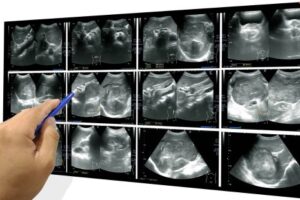
It is high-frequency sound that you cannot hear but it can transmitted and reflected in body. Reflected sound waves are detected by special device ( Transducer) to create images of body parts and structures. Now these Images are analyzed by radiologist to detect the various disease and structural abnormalities.
Because sound waves are used instead of radiation, so ultrasound scans are safe. It is painless and harmless for body.
3D/4D/5D Ultrasound (Sonography / USG)
UAD welcomes you for all kind of ultrasounds with world’s most advanced high end 4D/ 5D ultrasound machine providing 3D structure of internal organs in motion state, for better characterization of your diseases and fetal anomalies. We provide a wonderful experience of pregnancy USG in a comfortable and hygiene environment, where you may see your baby’s beautiful face, heart movements, kicking and yawning in realistic view on a large screen. In addition to regular USGs, all kinds of Doppler Studies & Liver Elastography -Fibroscan (2D-shearwave technology) suggesting liver diseases are undertaken at AUD.
All the USG will be performed by Dr. Bhoopchand Maurya ( MBBS, MD Radio-diagnosis, Consultant radiologist & Interventionologist ) who is highly experienced in pregnancy & infertility related ultrasounds, Color Doppler Echocardiography and interventional procedures ( Like- FNAC , Biopsy, Pigtail Insertion, PCN , CVS etc).
Our procedures meet world-class standards and PNDT guidelines. According to the PC-PNDT Act, pregnancy ultrasound are prohibited without a valid prescription, having the doctor’s UPMC/MCI registration number, phone number and other required details.
Book Your Free Consultation Call Today!
Get personalized guidance from our experts with a free consultation call. Don’t miss this chance to explore tailored solutions for your needs!
Other Service
At AUD all types Ultrasound & Color Doppler Scan are performed including-
| Obstetrics & Gynecology | Color Doppler | High Frequency USG | & Other Ultrasound |
|---|---|---|---|
|
|
|
|
Most Commonly Booked Tests


The Benefits of Ultrasound at Universal Diagnostic:
Non-Invasive and Safe: Ultrasound is completely non-invasive, requiring no incisions or injections. Unlike other imaging techniques, such as X-rays and CT scans, ultrasound does not use ionizing radiation, making it safer for patients, including pregnant women and children.
Real-Time Imaging: Ultrasound provides real-time images, allowing healthcare professionals to observe the movement of organs and blood flow. This real-time capability is particularly useful in procedures like guiding needle biopsies or draining fluids.
Wide Range of Applications: Ultrasound is a versatile tool used in various medical specialties, from obstetrics and gynecology to cardiology and orthopedics. Its ability to visualize soft tissues, blood vessels, and organs makes it an invaluable diagnostic tool.
Affordability and Accessibility: Compared to other imaging modalities, ultrasound is more affordable and widely available. At Universal Diagnostic, we offer competitive pricing and convenient scheduling to ensure our patients have easy access to high-quality ultrasound services.
No Radiation Exposure: One of the biggest advantages of ultrasound is the absence of radiation exposure, making it a preferred choice for imaging, especially for pregnant women and young children. This ensures the safety of both the patient and the developing fetus during obstetric ultrasounds.
Comfortable and Painless: The ultrasound procedure is comfortable and painless for the patient. The transducer is gently moved across the skin, and patients usually experience no discomfort during the examination.
Book your ultrasound at Universal Advanced Diagnostic today for a safe and quick health check!
Discover the power of non-invasive imaging with ultrasound—schedule your appointment at Universal Advanced Diagnostic today for safe, accurate, and real-time insights into your health!
How Does Ultrasound Work?
Ultrasound technology operates by emitting sound waves into the body using a device called a transducer. These sound waves bounce off tissues and organs, creating echoes that are captured and translated into images by the ultrasound machine. These images provide a real-time view of the inside of the body, helping healthcare professionals assess and diagnose a wide range of conditions.
The Transducer:
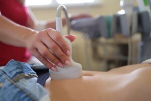
The ultrasound procedure begins with a device called a transducer, which emits high-frequency sound waves. The transducer is placed on the skin, usually after a gel is applied to ensure good contact. This gel helps transmit the sound waves more effectively into the body.
Sound Wave Transmission:
The transducer sends sound waves into the body, which then travel through tissues, organs, and fluids. When these sound waves encounter different types of tissues (such as muscle, bone, or fluid), they bounce back or “echo.”
Echo Reception:
The transducer also acts as a receiver, picking up the echoes that bounce back from the tissues. Different tissues reflect sound waves differently, creating variations in the echoes. For example, denser tissues like bone reflect more sound waves, while fluids like blood or amniotic fluid allow sound waves to pass through with little reflection.
Image Creation:
The ultrasound machine processes these returning echoes and converts them into real-time images. These images are displayed on a monitor, allowing healthcare professionals to visualize and assess the internal structures. The images can show the shape, size, and movement of organs, as well as blood flow and other important details.
Ultrasound is a versatile tool because it provides real-time imaging, allowing doctors to observe movement within the body, such as the beating of the heart or the movement of a fetus. It’s widely used because it’s safe, non-invasive, and does not involve radiation, making it suitable for various diagnostic purposes.


